

 Something else? Ask Aunt Vadge – bottom left of your screen!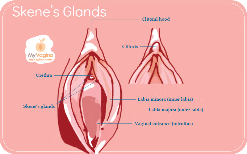

Skene’s glands are small paraurethral glands that sit around the lower end of the urethra, on the anterior (front) wall of the vagina. They are increasingly called the female prostate, because they share embryological origins with the male prostate and can produce prostate-specific antigen (PSA)1.
You might also see them called the lesser vestibular, periurethral or paraurethral glands. Whatever the name, they drain through the Skene’s ducts near the urethral opening, and the surrounding tissue – which includes part of the clitoris extending up into the vagina – swells during arousal, when the glands release a small amount of white fluid2.
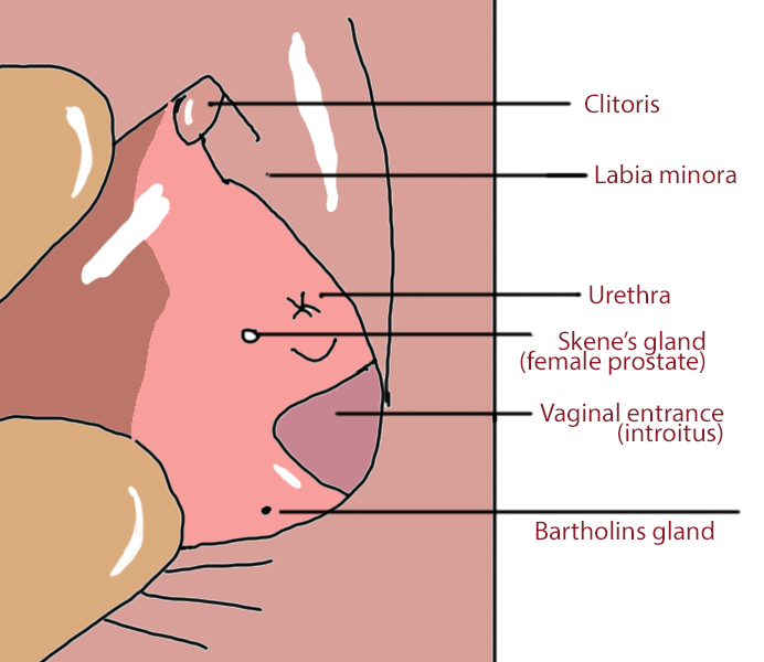
What Skene’s glands are and where they sit
Most of the time the Skene’s glands are small, not easily visible, and cause no symptoms. Their position, tucked right against the urethra, is why any problem here tends to look like a urinary or vaginal-wall issue – a cyst pressing on the urethra can change how you wee, and a rare tumour can mimic a urethral diverticulum.
Oddly, most diagrams of female anatomy leave the Skene’s ducts out altogether, and in some women the glands are so small they are barely there – so you could be forgiven for thinking they don’t exist at all.
Skene’s glands vs Bartholin glands
The Skene’s glands are often mixed up with the Bartholin glands, but they sit in different places and do different jobs.
Skene’s glands are at the front, around the urethra on the anterior vaginal wall, and make the prostate-like fluid described above. The Bartholin glands – also called the greater vestibular glands – sit further back, one on each side of the vaginal opening, and make mucus that helps with lubrication.
Both can block and form a cyst. A Skene’s cyst shows up near the urethra at the front, while a Bartholin cyst shows up at the back of the vaginal opening.
The female prostate: fluid, PSA and ‘squirting’
In some women, this female prostatic fluid leaves the urethra during sexual stimulation. The viscous white secretion is very similar in make-up to male prostatic fluid, including particular proteins and enzymes (Human Protein 1 and PDE5).
It has lower levels of creatinine but raised levels of prostate-specific antigen, prostatic acid phosphatase and glucose. Looked at closely, the male prostate and the Skene’s glands work so similarly that researchers increasingly treat the Skene’s glands as the female prostate.
This has had some unexpected consequences. In the past, PSA found in sexual-assault cases was treated as proof of male involvement – at a time when it was assumed women had no prostate and could not produce PSA. That assumption may have contributed to wrongful convictions3.
Does the fluid protect against UTIs?
There is a theory that these secretions are protective. Researchers have suggested that fluid deposited into the urethra during ejaculation might make a woman less likely to develop a urinary tract infection (UTI), particularly a sex-induced one4.
Female ejaculation (‘squirting’)
As a rough rule, the larger the Skene’s glands, the more likely someone is to be a ‘squirter’. Female ejaculation is complicated, though – not everyone with a vagina can do it, and some can’t help it. The small white secretion described above isn’t quite the same as the larger gush of watery fluid people describe during squirting.
The long-running debate about whether female ejaculation even exists was taken up in Emanuele Jannini’s 2002 work, which found highly variable Skene’s gland anatomy, sometimes absent entirely. If the glands drive female ejaculation and so-called ‘g-spot orgasms’, that variation may be why some people have no idea what the fuss is about.
History and discovery
The anatomist Regnier de Graaf described the female prostate as far back as 1672. In 1880, Alexander Skene drew attention to the glands and ducts again, which is how they came to carry his name. The shift now is back towards ‘female prostate’, as the more accurate term.
When the Skene’s glands cause problems
Most Skene’s glands never cause any trouble. When they do, it is usually a benign cyst; cancer here is genuinely rare. The glands have also been studied as a possible reservoir for HIV5.
Skene’s gland cysts: causes, signs and treatment
Skene’s gland cysts are far more common than cancer. They form when the duct becomes blocked, often after inflammation or infection.
Common symptoms of Skene’s gland cysts
- A smooth lump at the urethral opening that may change in size
- Pressure, discomfort or pain with sex or sitting
- Urinary urgency, dribbling, or a stop–start stream
- A visible swelling at the front of the vaginal opening
How Skene’s gland cysts are treated
Most Skene’s gland cysts are benign and straightforward to manage. A doctor will suggest treatment based on size, symptoms, and whether an infection is present.
- Watchful waiting – small, painless cysts can simply be monitored if they are not causing problems
- Targeted antibiotics – if the cyst is infected or forms an abscess, cultures can guide the choice; antibiotics alone rarely clear a blocked duct without drainage
- Drainage or marsupialisation – for painful, persistent or recurrent cysts or abscesses, opening the duct to create a drainage pathway is effective
- Excision – surgical removal of the cyst or involved duct is considered if cysts keep coming back
- Imaging or biopsy – if the lump is firm, fixed, ulcerated, rapidly enlarging or otherwise unusual, further imaging and biopsy may be recommended
- Aftercare and recurrence – sitz baths, rest and good hygiene aid recovery; report any new swelling, fever or ongoing urinary symptoms, as recurrence can happen and is treatable
Skene’s gland cancer: symptoms, risk, diagnosis and treatment
Skene’s gland cancer is extremely rare, with fewer than a few dozen cases reported worldwide6–9. Because of its rarity, it is often misdiagnosed as a urinary tract infection, a urethral diverticulum, or a vaginal-wall cyst.
Typical symptoms
- A persistent lump or swelling near the urethra
- Pain or pressure in the anterior (front) vaginal wall
- Blood in the urine or unusual discharge
- Urinary urgency, frequency, or difficulty emptying the bladder
- Recurrent urinary tract infections that do not respond to treatment
Risk factors
There are no proven risk factors for Skene’s gland cancer. Most reported cases have occurred in women over the age of 40.
Because the Skene’s glands are related to the prostate6, some tumours behave similarly, including producing PSA. That similarity is why a few cases have responded to hormone-based therapies, though the evidence is limited.
Diagnosis
Assessment starts with a doctor taking a thorough history and examining the area. If a lump is found, tests may include:
- Urine tests and swabs to rule out infection
- Pelvic MRI to work out whether the lesion is a cyst, abscess, diverticulum or tumour
- Cystoscopy or urethroscopy to look inside the urethra
- Biopsy to confirm the diagnosis and determine the type of tumour
- PSA testing in some cases, since Skene’s tissue can produce PSA
Getting the diagnosis right is what separates a benign cyst from a malignancy and points to the correct treatment.
Treatment options
Because so few cases exist, there is no single standard pathway. Reported treatments include:
- Surgical removal of the tumour
- Radiation therapy, either alone or after surgery
- Chemotherapy in selected situations
- Hormonal (androgen-deprivation) therapy in tumours that express PSA or resemble prostate adenocarcinomas
Prognosis depends on the stage at diagnosis and the tumour biology. Reliable survival statistics are not available given how few cases are documented, but early recognition and treatment give the best chance of a good outcome.
What to do if you notice symptoms
Skene’s glands are normal structures. Problems are uncommon and cancer is very rare – most issues are benign cysts. Even so, ongoing or unusual symptoms are worth checking.
If symptoms persist, get assessed by a doctor experienced in this area. An accurate diagnosis is the key to effective treatment and peace of mind.
Skene’s glands FAQ
Where are Skene’s glands located?
They are paraurethral glands located around the lower end of the urethra on the anterior vaginal wall. See our Skene’s glands anatomy diagram and Skene’s gland location in females for more detail.
What are common Skene’s gland cancer symptoms?
A persistent lump near the urethra, pain or pressure in the vaginal wall, urinary changes such as urgency or frequency, blood in urine, or recurrent UTIs that do not improve with antibiotics.
How rare is Skene’s gland cancer?
It is extremely rare, with fewer than a few dozen cases reported worldwide. Most lumps in this area are benign, such as Skene’s gland cysts.
How do doctors diagnose Skene’s gland problems?
Evaluation may include a medical history and examination, urine tests and swabs, pelvic MRI to define the lesion, cystoscopy or urethroscopy to inspect the urethra, and a biopsy of any suspicious tissue. PSA testing may be considered because Skene’s tissue can produce PSA.
What is the difference between a Skene’s gland cyst and cancer?
A Skene’s gland cyst is a blocked duct and is usually benign. Cancer is malignant and very rare. Imaging and biopsy are required to distinguish cysts from tumours.
How are Skene’s gland cysts treated?
Options include monitoring small cysts, antibiotics for infection, drainage or marsupialisation for persistent cysts, excision if cysts recur, and imaging or biopsy if the lump has atypical features.
What treatments are used for Skene’s gland cancer?
Treatment may involve surgical excision, radiation therapy, chemotherapy in selected cases, and hormone therapy for tumours that behave like prostate-type cancers. The plan depends on tumour type, stage, and individual factors.
When should I see a doctor?
Seek medical assessment if you notice a persistent lump near the urethra, urinary changes, unexplained bleeding, pain or pressure that does not improve, or recurrent urinary infections.
Are Skene’s glands the same as the female prostate?
Yes. The Skene’s glands are increasingly called the female prostate, because they share their origins with the male prostate and can produce prostate-specific antigen (PSA). You might also see them called the paraurethral or periurethral glands.
What is the difference between Skene’s glands and Bartholin glands?
Position and job. The Skene’s glands sit at the front, around the urethra, and make a prostate-like fluid. The Bartholin glands sit further back, one on each side of the vaginal opening, and make mucus for lubrication. Both can form a cyst.
- Zaviačič M, Ablin RJ. The Female Prostate. JNCI: Journal of the National Cancer Institute. 1998;90(9):713–713.
- Dagur G, Warren K, Imhof R, Gonka J, Suh Y, Khan SA. Clinical implications of the forgotten Skene's glands: A review of current literature. Polish Annals of Medicine. 2016;23(2):182–190.
- Longo VJ. The female prostate. Urology. 1982;20(1):108–109.
- Moalem S, Reidenberg JS. Does female ejaculation serve an antimicrobial purpose?. Medical Hypotheses. 2009;73(6):1069–1071.
- Ablin RJ. HIV-Related Protein in the Prostate: A Possible Reservoir of Virus. American Journal of Clinical Pathology. 1991;95(5):579–579.
- Kaufman ME, Miller DT, Ullah A, et al. Skene's Gland Adenocarcinoma: Borrowing From Prostate Cancer Experience for the Evaluation and Management of a Rare Malignancy. Urology. 2021;151:182–187.
- Kyriazis G, Varughese A, Rodrigues G, Simms M. A Rare Case of Skene’s Gland Adenocarcinoma. Clinical Genitourinary Cancer. 2020;18(3):e300–e302.
- Slopnick EA, Bagby C, Mahran A, et al. Skene's Gland Malignancy: A Case Report and Systematic Review. Urology. 2022;165:36–43.
- Tregnago AC, Epstein JI. Skene’s Glands Adenocarcinoma. American Journal of Surgical Pathology. 2018;42(11):1513–1521.