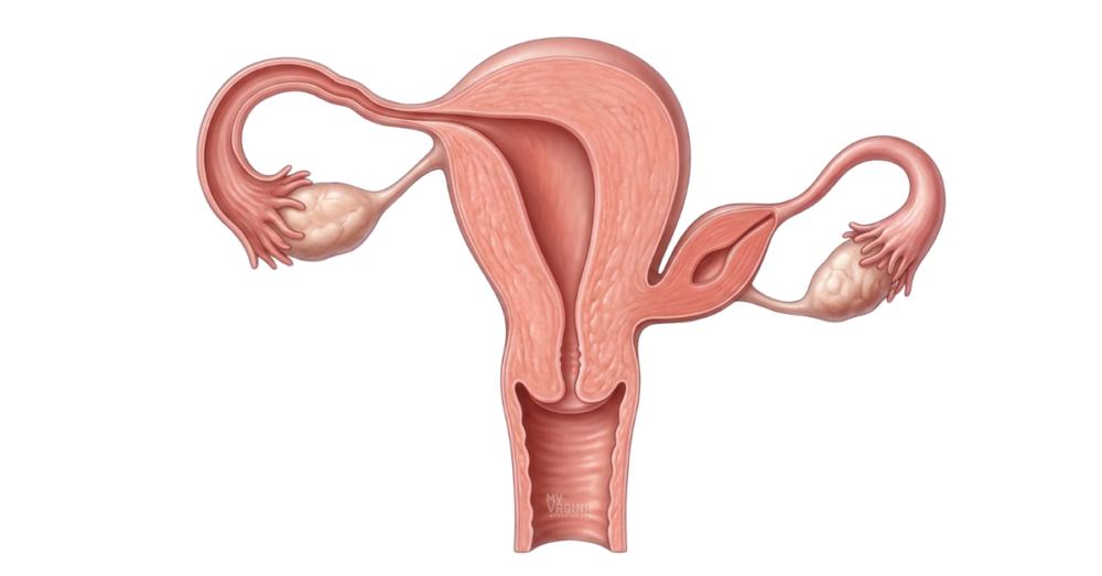

An obstructed uterine horn is a difference in the shape of the uterus that you are born with, where one side forms as a separate, partly or fully closed-off chamber – a rudimentary horn – that does not drain properly into the main uterus and vagina. It is a type of Müllerian duct anomaly, and it most often sits alongside a one-sided (unicornuate) uterus.1–2
The two things that matter most are pain and pregnancy. If the horn has a working endometrial lining, menstrual blood is made each month but has nowhere to go, which causes cyclic, often one-sided pelvic pain. And in the rare event a pregnancy implants in the horn, it can be dangerous. Treatment is surgical, and this is firmly a doctor’s area, not a naturopath’s.2–4
What is an obstructed or rudimentary uterine horn?
Your uterus normally forms when two tubes, the Müllerian (paramesonephric) ducts, fuse together in early development. When one duct only partly develops, you can end up with a single main uterus on one side and a smaller, underdeveloped horn on the other. That smaller horn is called rudimentary.1
A rudimentary horn can be solid, or it can have a small cavity lined with endometrium. It is called obstructed or non-communicating when that cavity does not connect to the main uterine cavity or the cervix, so anything it produces is trapped. This is the version that tends to cause problems. In the international classification of these differences, a one-sided uterus with a functional horn is grouped as a hemi-uterus with a rudimentary cavity.1–2
It often travels with related anatomy. The kidney on the same side as the rudimentary horn is sometimes missing or sits in an unusual place, because the kidneys and reproductive tract develop side by side.2 An obstructed horn is closely related to the unicornuate uterus, and sits in the same family as other Müllerian differences such as uterus didelphys and uterine hypoplasia.
What are the symptoms?
Many people with a uterine horn have no symptoms at all, particularly if the horn is solid or does not have a working lining. When symptoms do appear, they usually start within a few years of the first period, as the trapped lining begins to bleed each month with nowhere to drain.3
The most common signs are:
- Severe period pain, often worse on one side
- Cyclic pelvic pain that builds month on month
- A tender lump or mass that can be felt in the lower abdomen
- Pain that does not settle with the usual period-pain measures
The trapped blood can build up inside the horn (a haematometra) and, over time, push back along the fallopian tube, which is one reason this anomaly is linked with period pain and endometriosis on the affected side.2–3
Why does it happen?
An obstructed uterine horn is a developmental difference, not something you did or could have prevented. It happens when the Müllerian ducts do not fuse and canalise in the usual way while you are still a developing foetus.1
There is no single known cause, and in most cases it is not inherited in any predictable way. Because it forms before birth, it is present from the start, even though it may not be picked up until the teenage years or later, or sometimes only during a fertility check or a scan for another reason.3
The serious risk: pregnancy in a rudimentary horn
This is the part that makes early diagnosis worth chasing. A pregnancy can, rarely, implant inside a rudimentary horn. Surprisingly, this can happen even when the horn is non-communicating, because sperm can travel across the pelvis from the other side to reach the egg – what is known as transperitoneal migration.2
A horn is not built to carry a pregnancy. Its muscular wall is thin and does not stretch well, so a pregnancy growing there carries a high risk of rupture, often before 20 weeks, which is a medical emergency. Pregnancies in a rudimentary horn are uncommon, but because the consequences are severe, knowing the horn is there beforehand changes everything about how a pregnancy is monitored.2–3
How is it diagnosed?
Diagnosis usually starts with an ultrasound, which can show a one-sided uterus and a separate horn. The picture is not always clear, though, and a rudimentary horn is easy to mistake for a fibroid or an ovarian mass.3,5
Magnetic resonance imaging (MRI) is the most useful test, because it shows the soft tissue in detail and can tell whether the horn has a working lining, whether it connects to the main cavity, and how it relates to the cervix.5 Imaging of the kidneys is often done at the same time, since a missing or misplaced kidney is a common companion finding.2
How is it treated?
When an obstructed horn with a functional lining is causing pain, or carries a pregnancy risk, the usual treatment is surgical removal of the horn, commonly by keyhole (laparoscopic) surgery.4 Removing it relieves the cyclic pain, stops blood building up, and takes away the risk of a future pregnancy implanting there.3–4
A solid horn with no lining and no symptoms may simply be monitored rather than removed. Each situation is assessed individually, and the plan depends on whether the horn has a cavity, whether it is causing pain, and what someone wants for future fertility.4
In our experience, it is the pain that does not quite fit – one-sided, building over time, not eased by the usual measures – that should prompt a proper look at the underlying anatomy rather than another round of period-pain advice.
This is a doctor’s job, not a naturopath’s. An obstructed horn is a structural problem that needs a GP or gynaecologist, imaging and, usually, surgery. We rarely see it in our clinic, and almost never in the teenagers who most often present with it – by the time someone reaches us, the diagnosis and surgery are well behind them.
Frequently asked questions
Can you still get pregnant with an obstructed uterine horn?
Often, yes. Most people with a rudimentary horn also have a functioning main uterus on the other side, and pregnancies in that main uterus can go well. The concern is specifically a pregnancy implanting in the horn itself, which is why diagnosis matters.2
Is an obstructed uterine horn the same as a unicornuate uterus?
They are closely linked but not identical. A unicornuate uterus is a single-sided uterus; a rudimentary horn is the smaller, underdeveloped second side that may or may not be present alongside it.1–2
Why does it cause one-sided pain?
If the horn has a lining, it sheds blood each month like the rest of the uterus, but the blood is trapped because the horn does not drain. That build-up causes cramping and pressure on the affected side.3
Could it affect my kidneys?
The kidney on the same side is sometimes missing or sits in an unusual position, because the urinary and reproductive systems form together. This is why doctors often image the kidneys when they find a uterine horn.2
Will surgery affect my fertility?
Removing a rudimentary horn does not remove your main uterus or your ovaries, so the goal is to relieve pain and reduce risk while protecting future fertility. The details depend on your own anatomy, which a gynaecologist will explain.4
What to do next
If you have severe, one-sided or worsening period pain, or you have been told you have a uterine horn or a one-sided uterus, the right next step is a GP or gynaecologist who can arrange proper imaging, ideally including an MRI. This is a structural diagnosis that needs a doctor.
If period pain has been part of a longer story of irregular or absent periods, our guide on reasons you don’t have your period yet is a useful read alongside that medical work-up.
If you would like help making sense of your symptoms before an appointment, you can chat with Aunt Vadge’s Assistant in the bottom left of your screen, or book in with one of our practitioners for general support around your gynaecological health.
This article is general information, not a substitute for personalised medical advice. An obstructed uterine horn needs assessment and management by a qualified doctor.
- Grimbizis GF, Gordts S, Di Spiezio Sardo A, et al. The ESHRE/ESGE consensus on the classification of female genital tract congenital anomalies. Hum Reprod. 2013;28(8):2032-2044.
- Reichman D, Laufer MR, Robinson BK. Pregnancy outcomes in unicornuate uteri: a review. Fertil Steril. 2009;91(5):1886-1894.
- Jayasinghe Y, Rane A, Stalewski H, Grover S. The presentation and early diagnosis of the rudimentary uterine horn. Obstet Gynecol. 2005;105(6):1456-1467.
- Letterie GS. Management of congenital uterine abnormalities. Reprod Biomed Online. 2011;23(1):40-52.
- Chandler TM, Machan LS, Cooperberg PL, Harris AC, Chang SD. Müllerian duct anomalies: from diagnosis to intervention. Br J Radiol. 2009;82(984):1034-1042.