
A hysteroscopy is one of the few tests that lets a doctor look directly inside your uterus, rather than guessing from the outside. A thin telescope with a camera on the end is passed gently through the vagina and cervix into the uterine cavity, and the view is shown on a screen.1
It is the reference test for assessing the inside of the uterus, and it has quietly become something most people can have while awake, in a clinic room, in a matter of minutes.1 If you have been told you need one, here is what it is for, what it actually feels like, and what to expect before, during and afterwards.
What is a hysteroscopy?
A hysteroscopy is the inspection of the uterine cavity using a hysteroscope – a narrow tube, often only a few millimetres wide, carrying a light and a camera. The cervix is the natural doorway in, so no cuts are needed.1
To open the cavity enough to see, the uterus is gently distended with fluid (usually saline) or, less commonly, carbon dioxide gas. The walls separate slightly, and the lining, the openings of the fallopian tubes and any growths come into clear view.1
Modern outpatient hysteroscopy often uses a ‘no-touch’ approach called vaginoscopy, where the scope is guided in without a speculum and without gripping the cervix. This is now the recommended standard technique because it is more comfortable for most people.1
Why might you need a hysteroscopy?
Most hysteroscopies are arranged to find the cause of abnormal bleeding, or to investigate something already seen on an ultrasound. It is used to look for and, increasingly, to treat a range of issues in the same sitting.1
Common reasons include:
- Heavy periods or bleeding that does not fit your usual pattern
- Bleeding between periods or after sex, and any bleeding after menopause
- Investigating polyps or fibroids that bulge into the cavity
- Checking the lining when there is concern about endometrial hyperplasia, endometrial cancer or other uterine cancers
- Fertility investigations, recurrent miscarriage, or scar tissue and structural differences inside the uterus
- Removing a stuck or hard-to-find intrauterine device (IUD), or retained tissue after a pregnancy
A hysteroscopy is one piece of the picture, not the whole story. It often follows a pelvic exam and a transvaginal ultrasound, and sits alongside other tests in the wider workup for the uterus and ovaries.1
Diagnostic and operative hysteroscopy
There are two overlapping jobs a hysteroscopy can do. A diagnostic hysteroscopy is purely a look-and-see: the doctor inspects the cavity and, if needed, takes a small sample of the lining (a biopsy).1
An operative hysteroscopy goes a step further, passing fine instruments down the same scope to remove a polyp, shave away a fibroid, divide scar tissue, or take out a foreign body.1
Where the problem is small and clearly visible, many clinics now offer a ‘see-and-treat’ service – finding and fixing the issue in one outpatient appointment, rather than booking a separate operation.1 Whether this is suitable depends on the size and type of growth, your comfort, and the equipment available.
Where it is done, and whether you are awake
Hysteroscopy can be done two ways. The first is an outpatient or office procedure, while you are awake, with no anaesthetic or only a local one. The second is in an operating theatre under general or regional anaesthetic.2
The shift in recent years has been firmly towards the awake, outpatient route, supported by good counselling and a calm, well-staffed clinic set-up.2 It avoids a general anaesthetic, lets you go home quickly, and for diagnostic work it is just as informative.
A theatre procedure is generally chosen for larger or more complex operative work, if a previous awake attempt was too uncomfortable, or where access through the cervix is difficult. An example is cervical stenosis, a narrowing of the cervical canal.1 The choice is yours to make with your clinician, and it is reasonable to ask why one setting is being recommended over the other.
What actually happens during the procedure
Before you go in
An outpatient hysteroscopy is usually booked when you are not bleeding heavily, as a clear view is easier mid-cycle. You may be advised to take a simple pain reliever an hour beforehand, and you can normally eat and drink as usual.1
You will be asked to empty your bladder, then to lie back with your legs supported. Good clinics talk you through each step and check in with you as they go – this is part of keeping the experience comfortable, and that matters.2
During the procedure
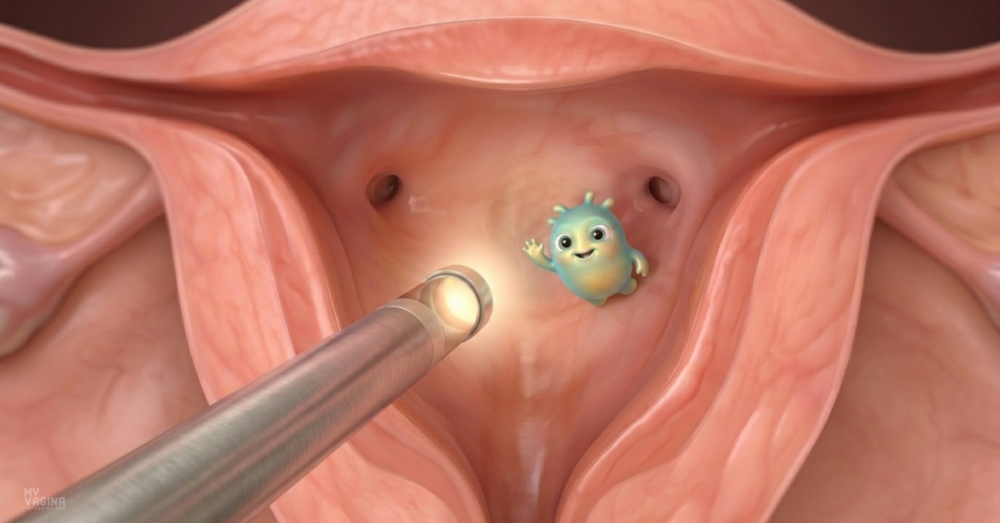
With the vaginoscopy approach, the scope is passed gently along the vagina, through the cervix and into the uterus, using fluid to open the way. There is often a feeling of pressure or period-like cramping as the cervix is crossed and the cavity fills.1
The doctor inspects the lining and the tubal openings, and may take photos. If a biopsy or a minor procedure is planned, it happens now. A diagnostic look often takes only a few minutes; operative work takes a little longer.1
Afterwards
After an outpatient hysteroscopy most people rest for a short while, then go home the same day, often without needing anyone to collect them. Expect some cramping and light bleeding or spotting for a few days, which a regular pain reliever usually handles.1
If you had a general anaesthetic, you will need someone to take you home and stay with you, and you should not drive for the rest of the day. It is sensible to use pads rather than tampons or a menstrual cup while you are spotting, and to ask your clinic when sex and swimming are fine again.
Does a hysteroscopy hurt?
This is the question almost everyone wants answered, and the honest reply is that it varies. Many people feel only mild, period-like cramping and find an awake procedure very manageable; for others it is properly painful, and that experience is valid and worth planning for.1
Pain tends to be greater when the cervix is tight, when there has not been a vaginal birth, and with longer or operative procedures. The vaginoscopy ‘no-touch’ technique helps, because gripping the cervix is one of the more uncomfortable steps.1
Where extra pain relief is needed, injecting local anaesthetic around the cervix is the most effective option studied, and your clinic should discuss what is available before you start.3 You are allowed to ask to pause or stop at any point, and to move to a theatre procedure under anaesthetic if an awake attempt is too much. Knowing this in advance takes a lot of the worry out of it.
Risks and complications
Hysteroscopy is considered very safe, and serious problems are uncommon. With a diagnostic procedure the most frequent event is a brief faint or light-headed feeling (a vasovagal reaction), which settles quickly with rest.4
The complication clinicians most watch for is a uterine perforation – a small hole made by an instrument passing through the uterine wall. In operative hysteroscopy this happens in roughly 1 in 100 procedures, and more often with the trickiest work such as dividing dense scar tissue; it is rarer still in simple diagnostic cases.4
Other recognised risks include infection, heavier bleeding, and, in longer operative cases, the body absorbing too much of the distension fluid – which is why clinics measure the fluid carefully throughout.4 Most perforations cause no lasting harm and are managed conservatively, but they are the reason an experienced operator and good monitoring matter.
How accurate is a hysteroscopy?
Because it gives a direct view, hysteroscopy is very good at spotting structural problems in the cavity. In people with abnormal bleeding, it picks up polyps with a sensitivity of around 95 per cent, and fibroids bulging into the cavity around 97 per cent.5
It is also highly specific for ruling out cancer, meaning a normal-looking cavity is reassuring. It is less reliable on its own for endometrial hyperplasia, where the lining can look normal but behave abnormally under the microscope.5
The camera shows the shape of the tissue, but not what is happening in the cells. So when hyperplasia or cancer is a concern, the camera view is paired with a biopsy, and the tissue diagnosis is what counts.5 A hysteroscopy that looks clear is not a substitute for sampling the lining when sampling is indicated.
What if your hysteroscopy comes back normal?
A normal result is common, and it is useful – it rules out polyps, fibroids in the cavity and other structural causes, which narrows the search considerably. But normal does not always mean nothing is wrong.
The cavity can look completely clear while the real trouble sits in the vaginal microbiome or in low-grade inflammation – neither of which a hysteroscope can see. This is exactly the group we tend to pick up in our clinical work. These are people told everything looked fine, who are still dealing with discharge, irritation, recurrent infections or symptoms that will not settle.
If that sounds like you, the useful next step is to look at what a structural test cannot assess. A thorough microbiome test shows whether protective bacteria have been displaced by disruptive bacteria, which points to a very different line of treatment than anything a hysteroscopy would prompt.
Frequently asked questions
How long does a hysteroscopy take?
A diagnostic look often takes only a few minutes. With a biopsy or a small operative step, allow a little longer, though the appointment itself is usually well under half an hour.1
Will I be put to sleep?
Not usually. Most diagnostic and many minor operative hysteroscopies are done while you are awake, with no anaesthetic or a local one. A general anaesthetic is kept for larger procedures or when an awake attempt is too uncomfortable.2
How should I prepare?
Follow your clinic’s instructions, take any recommended pain relief beforehand, and try to attend when you are not bleeding heavily. Bring a pad for afterwards, and ask in advance whether you will be awake or asleep so you can plan your day.1
What is the recovery like?
Mild cramping and light spotting for a few days is normal. Most people return to their usual routine within a day or two. Contact your clinic if you develop heavy bleeding, severe pain, a fever, or smelly discharge, as these can signal infection.1
How is it different from an ultrasound?
A transvaginal ultrasound uses sound waves to image the uterus and ovaries from outside the cavity, and is often the first test. A hysteroscopy looks directly inside the cavity and can take a sample or treat a problem at the same time, so the two are complementary rather than interchangeable.5
Can a hysteroscopy help with fertility?
It can. Polyps, fibroids in the cavity, scar tissue and some structural differences can affect implantation, and treating them hysteroscopically is part of a wider fertility workup. Scar tissue inside the uterus is also a recognised cause of absent or very light periods.1
Is a hysteroscopy the same as a hysterectomy?
No, and the similar names cause real confusion. A hysteroscopy looks inside the uterus and leaves it in place. A hysterectomy is major surgery to remove the uterus.
What to do next
If a hysteroscopy has been suggested, ask your clinician what they are looking for, whether it will be done awake or asleep, and whether anything can be treated at the same visit. Those three answers will tell you most of what you need to plan.
It also helps to understand the bigger picture. Abnormal bleeding has many possible drivers – from adenomyosis and endometriosis to hormonal causes – and a hysteroscopy is one tool among several. Our guide to diagnostic procedures for the vulva, vagina and reproductive organs walks through how these tests fit together.
If you would like to talk something through, Aunt Vadge’s Assistant is in the bottom left of your screen, and our practitioners are available for a proper one-on-one if you want personalised guidance.
This article is general information, not a substitute for individual medical advice. Please discuss your situation with your own clinician.
- De Silva PM, Smith PP, Cooper NAM, Clark TJ. Outpatient Hysteroscopy (Green-top Guideline No. 59). BJOG. 2024;131(13):e86–e110.
- Namkung J, Cho A, Koo YJ, et al. Clinical practice in office hysteroscopy. Obstet Gynecol Sci. 2025;68(3):175–185.
- Cooper NA, Khan KS, Clark TJ. Local anaesthesia for pain control during outpatient hysteroscopy: systematic review and meta-analysis. BMJ. 2010;340:c1130.
- Aas-Eng MK, Langebrekke A, Hudelist G. Complications in operative hysteroscopy – is prevention possible?. Acta Obstet Gynecol Scand. 2017;96(12):1399–1403.
- Gkrozou F, et al. Hysteroscopy in women with abnormal uterine bleeding: a meta-analysis on four major endometrial pathologies. Arch Gynecol Obstet. 2015;291:1347–1354.